ASPHERE
A High-Resolution k-Space Microscope
Here we present our recently developed photoelectron spectrometer ASPHERE (Angular Spectrometer for Photoelectrons with High Energy REsolution), which has proven to be a very versatile instrument for high-resolution ARPES work. ASPHERE is now part of our experimental end station at the HONORMI beamline (see Fig.1) at the Hamburger Synchronstrahlungslabor, but the entire experimental setup is also compatible with beamline BW3. The major characteristics of the photoemission experiment are:
- high energy resolution (10 meV) in consequence of complete fringe field compensation
- tuneable angular resolution (0.2° - 5°) by means of an iris aperture
- rotating analyzer (fixed crystal) technique with automated high-precision (0.2°) angle scanning
- good count rates due to multidetection system
- synchrotron radiation in a wide energy range (HONORMI: 5 - 30 eV; BW3: 40 - 1500 eV)
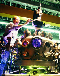
FIG. 1. The ASPHERE experiment at HASYLAB.
Spectrometer Design
In the construction of ASPHERE we have chosen the concept of a large 180° spherical analyzer with a 4-element electrostatic input lens used for retarding and focusing. The central radius of the condenser is 100 mm and the gap between the hemispheres is 30 mm. The fringe field of the condenser has been corrected for by slightly tilting the input lens against the base plane of the hemispheres. The geometry of the input lens allows for an uniform magnification of 1 over a wide retardation-ratio range and the angular acceptance can be varied continuously from 0.2° to 5° by means of an iris aperture. To improve the speed of data acquisition three standard channeltron detectors have been installed in the image plane of the analyzer, but the analyzer is also compatible with a channelplate multidetection system. In order to be able to sweep the analyzer across the emission hemisphere we have constructed a two-axes goniometer with computer-controlled stepper motors. In our setup, the working distance to the sample position is 50 mm leaving enough space for further experimental probes. The complete ASPHERE spectrometer is shown in Fig. 2.
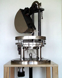
FIG. 2. ASPHERE mounted on the two-axis goniometer.
Energy Resolution
Giving proof of ASPHERE's high energy resolution, Fig. 3 depicts a gold Fermi edge measured at 9 eV photon energy and a temperature of 10 K. The data were numerically fitted (solid blue line) by assuming a linearly increasing density of states which is first cut off by the Fermi-Dirac distribution and then convoluted with a Gaussian spectrometer response function. The fitted overall energy resolution is 10 meV including a contribution of 6 meV from the exciting photons.
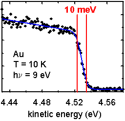
FIG. 3. Gold Fermi edge showing an energy resolution of 10 meV.
Photoelectron Angular Distributions
However, the main quality of ASPHERE is high-precision k-space mapping using the rotating analyzer technique, i.e. during the photoemission measurements the light incidence is always at fixed angle relative to the sample with the light polarization residing fixed inside the crystal. In Figs. 4 and 5 we exemplarily present some photoelectron angular distributions that were taken from different transition metal dichalcogenides for various photon and electron energies. By imaging the momentum-resolved valence band structures including the different topologies of the Fermi surface in great detail, these angle-scanned measurements directly uncover the beauty of electronic structures within the first and higher Brillouin zones of k-space. Note that no data manipulation such as smoothing, normalization or background correction has been applied to the photoemission images shown below.
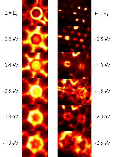
FIG. 4. Photoelectron angular distributions from NbSe2 (left: hν = 24.5 eV) and TiSe2 (right: hν = 80 eV).
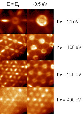
FIG. 5. Photoelectron angular distribution from TiTe2.
Specifications
Angular acceptance: ±0.2° - ±5°
3 Channel detection system
Mean radius: 100 mm
Entrance / exit slits: 1 mm
4 Lens elements
Lens magnification: 1
Working distance: 50 mm
- horizontal: 0° - 355°
- vertical: -70° - +30°
Angular precision:
- horizontal: 0.1°
- vertical: 0.25°
Closest approach angle: 20°
Stepper motor control unit
Precision: ±2 meV
Noise: <2 mV
Pass energy range: 0.5 - 50 eV
(continuously adjustable)
Kinetic energy range:
- UPS: 0 - 100 eV
- XPS: 0 - 1.5 keV
Reference

Contact Information
ALS, Berkeley
E-Mail: KRossnagel@lbl.gov
IEAP, University of Kiel
E-Mail: harm@physik.uni-kiel.de
IEAP, University of Kiel
E-Mail: kipp@physik.uni-kiel.de